DOE-BER grant seeks new imaging approaches to observe plant-microbe interactions
Michigan State University and the Department of Energy's SLAC National Accelerator Laboratory, operated by Stanford University, are working to build new microscopes that allow scientists to peer into plant cells like never before.

Federica Brandizzi, MSU Foundation Professor of plant biology. Credit: Derrick Turner, University Communications
Using a three-year, $507,264 grant from the Department of Energy Office (DOE) Office of Science’s Biological and Environmental Research (BER) program, MSU plant biologist Federica Brandizzi and her lab are collaborating with Stanford’s Soichi Wakatsuki, professor of photon science at the SLAC, professor of structural biology in the Stanford School of Medicine and the grant’s principal investigator, to create optical and X-ray multimodal-hybrid microscope systems for live imaging of plant stress responses and microbial interactions.
“These microscopes will have the speed and resolution to image single particles in biological systems,” said Brandizzi, an MSU Foundation Professor in the College of Natural Science’s Department of Plant Biology, an MSU-DOE Plant Research Laboratory faculty member, and grant co-principal investigator. “This is something that is very challenging to achieve.”
Brandizzi met Wakatsuki at a virtual DOE workshop during the pandemic. Wakatsuki’s passion in building new and powerful microscopes and his interest in the Brandizzi Lab’s ability to work in plant cell biology and microscopy led him to the belief that the two labs could create something great.

“This is a truly multidisciplinary effort to develop powerful X-ray, fluorescent microscopy and cryo-electron microscopy and tomography imaging systems,” Wakatsuki said. “The collaboration with Brandizzi’s group is vital to ensuring these new imaging tools work as expected during complex experiments, specifically visualization of membrane trafficking in plants’ defense against pathogens.”
SLAC is the central hub of this collaboration with MSU and Stanford University and will eventually host the experiments at the Stanford Synchrotron Radiation Lightsource (SSRL). The project is known as Fluorescence Lifetime Imaging Microscopy (FLIM) and it builds on SLAC’s close collaboration with Mark Kasevich, a professor of physics at Stanford, whose group is developing ultrafast FLIM devices as part of another DOE BER funded project.
In this new project, SLAC will build another FLIM instrument and integrate it with hybrid X-ray/FLIM imaging and cryo-EM/ET. SLAC researchers will work with plant biologists Brandizzi and Mary Beth Mudget, the senior associate dean of Stanford’s Natural Sciences in the School of Humanities and Sciences Department, to image plant-pathogen interactions.
For its part, the Brandizzi lab will provide Arabidopsis thaliana—a small flowering plant widely used as a model organism in plant biology—to observe the interaction of these plants with microorganisms, specifically a fungal pathogen. The objective is to understand how the cell surface responds to the invasion of the pathogen.
Current constraints of technical equipment have hindered research on the response and speed of how a plant reacts to stressors, like pathogens. Two goals of the collaboration are to have a product in place that can answer these questions that will then lead to an understanding of which proteins will make a plant more resistant to pathogens.
This initiative is also made possible by the MSU Foundation, which has funded Bhandari’s position in the Brandizzi lab for the past few years.
“Deepak was immediately ready to use his expertise in plant pathogen interaction on this new grant,” Brandizzi said. “Without the investment from MSU and the MSU Foundation Professorships, generating data and securing expertise for this kind of exciting new collaborations would not be possible."
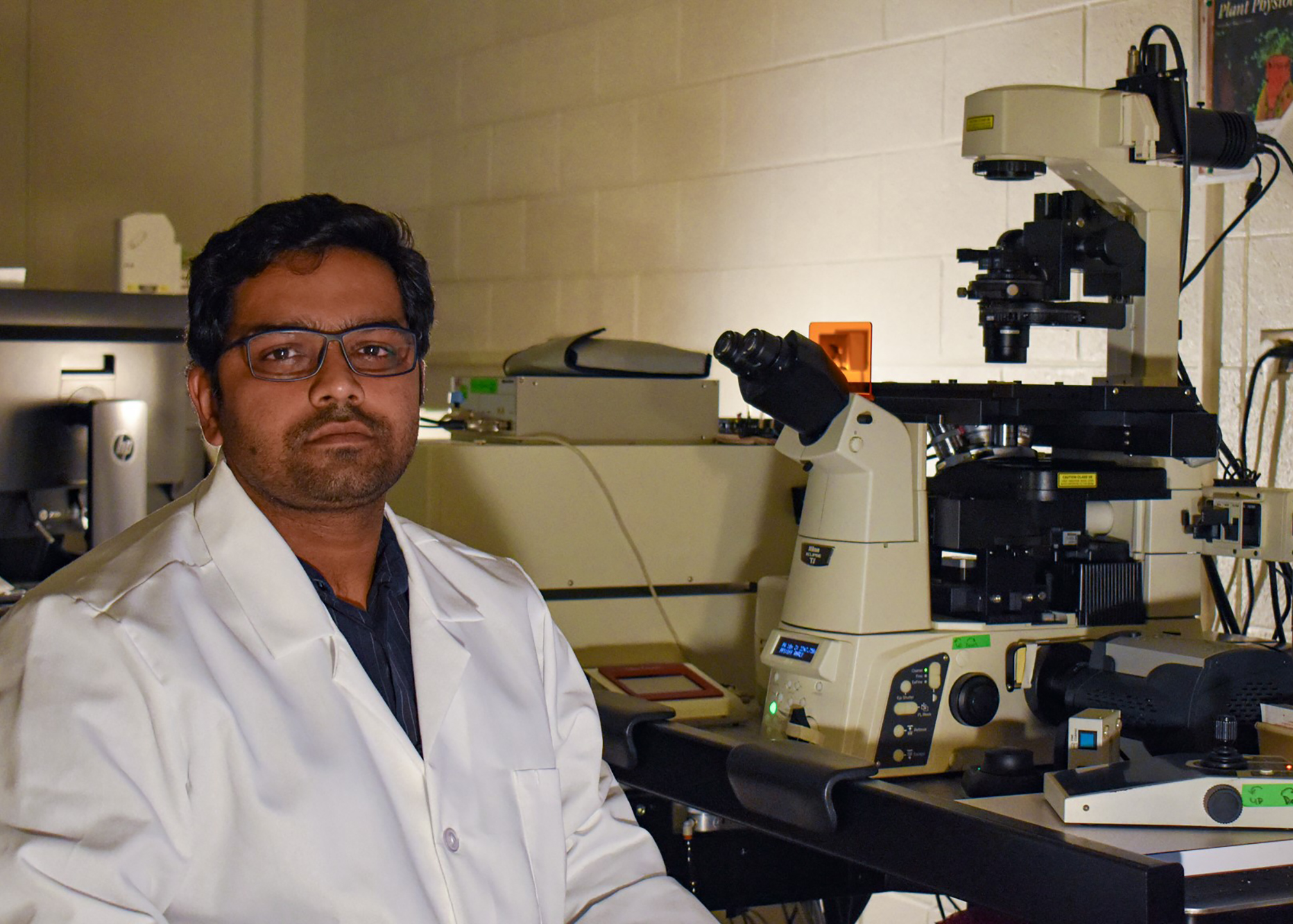
Banner image: Deepak Bhandari (above), a postdoc in the Brandizzi lab, uses microscopes
to study Arabidopsis thaliana for his research under this grant. The Brandizzi lab
will provide Arabidopsis thaliana (a common mustard plant) to observe the interactions
of these plants with microorganisms, specifically a fungal pathogen. The aim is to
understand how the cell surface responds to the invasion of the pathogen. Credit:
Kara Headley, MSU-DOE Plant Research Laboratory