MSU's Mark Reimers receives NIH grant to characterize interactions in the brain's core circuits
The Reimers lab at Michigan State University is reframing scientific understanding of how the brain works. To extend this research, MSU neuroscientist Mark Reimers, in collaboration with Ariel Gilad at the Hebrew University in Jerusalem, has been awarded a five-year, $1.2 million grant from the National Institutes of Health (NIH) under the Collaborative Research in Computational Neuroscience (CRCNS) grant program.
The CRCNS program is an international, multi-institution partnership that supports collaborative activities to advance the understanding of nervous system structure and function, mechanisms underlying nervous system disorders, and computational strategies used by the nervous system.
With a M.S. in computer science and a Ph.D. in probability theory, Reimers, an associate professor, joined MSU’s neuroscience program in the College of Natural Science in 2015. He works in the Institute for Quantitative Health Science and Engineering (IQ) and has a special interest in large-scale data analysis within computational biology.
“Even though our best tools for understanding brain function remain inadequate, gaining deeper insight to the brain is a compelling challenge.” Reimers said. “Over the past 50 years, most conventional neuroscience has focused on studying the brain, one area at a time, using the metaphor of information processing. We hope to shift the conversation toward a more dynamic, physiological view. If you record from the whole brain, you realize that it’s quite dynamic and interactive.”
Funding from the grant will go directly to Reimers’ research on understanding the interaction between the two largest classes of regions in the brain, the many nuclei of the thalamus and the many regions of cortex, which are in constant dialogue with each other. The goal of this project is to characterize the network circuitry of the brain and its dynamics in detail. For example, a sound enters the brain through the sensory thalamus, which sends signals to the sensory cortex, which then negotiates with several other thalamic regions to recruit other cortical regions to respond or ignore the sound.
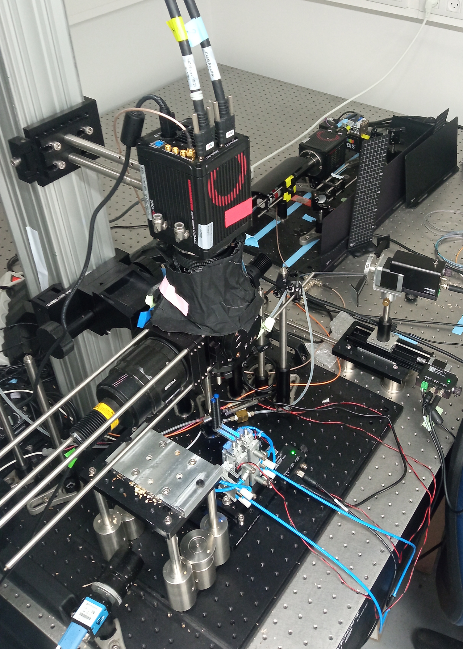
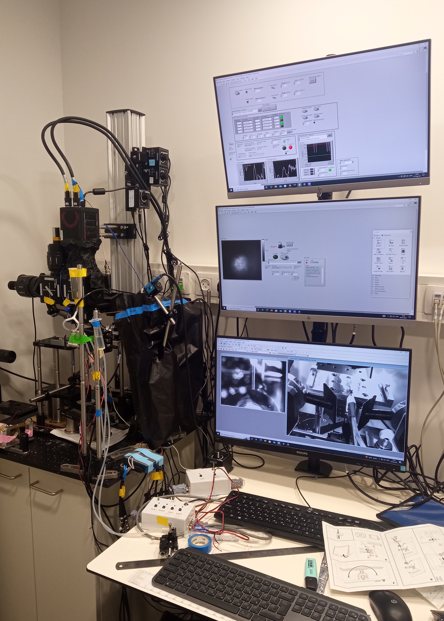
Reimers and Gilad will document how regions and circuits compete as the signals bounce back and forth several times per second. To capture dynamics from a large part of the brain, the team needs to record many areas of the brain simultaneously.
“What’s unique about this study is that it will take recordings from dozens of areas of the thalamus and dozens of areas the cortex simultaneously, recording neural activity fluctuations on time scales of less than half of a second,” Reimers said. “As far as we know, we are creating the first large-scale characterization of the communication within the tangled network at the core of consciousness at a ‘moment-by-moment’ sub-second time scale.”
By employing optogenetics, a method that uses light to activate or deactivate neurons, the team will start to uncover causality. Gilad sends his results to Reimers’ lab for analysis. The Reimers lab will characterize how often activity in one region of the brain is followed by activity in other regions, and Gilad lab tests these regions one at a time by stimulating or shutting down the nodes to record the changes, and especially to see if the circuit will reroute and if it does, how much longer it takes.
“The dataset produced in our experiments is rich and multidimensional,” Gilad said. “Mark brings very strong computational tools that can extract and quantify distinct network motifs within the broad thalamocortical networks. We are thrilled to be able to collaborate with him and learn to implement computational tools on our datasets.”
At the end of five years, they hope to have a fairly accurate map of dialogue between thalamic regions and cortical areas during many basic cognitive tasks, showing which connections are being used. This map may be important for treating psychiatric problems and neurological disorders. Learning how circuits in the brains of stroke patients reroute to other circuits to perform basic tasks could be important for stroke patients’ recovery. For psychiatric and neurological disorders such as Parkinson’s and ADHD, stimulating or shutting down nodes where symptoms occur could illuminate better treatments.
While they continue this research, Reimers and his team are developing a new imaging system that should be able to map 10 times as many neurons over about 10 times an area of the brain for less than half the cost of most systems. If they are successful, this would allow them to characterize their dynamics of communication among sub-networks in each area.
Banner image: A small section of a sample colormap figure representing brain activity in 45 different regions, together with three kinds of behavior, for one mouse during one test. Credit: Reimers lab